Histology 1

Mammalian Histology-B408
Department of Biological Sciences
University of Delaware
Mammalian Histology (B408) is taught in the fall semester annually and is one of the courses required for the Medical Scholars Program with the Jefferson Medical College. Consequently, it is taught at a comprehensive level and concentrates heavily on human tissues and organ systems. A strong component of this course is tissue structure at the ultrastructural level and how it relates to structure-functional relationships at the light microscopic level. A sizeable collection of color light microscopic images as well as black and white electron microscopic images have been archived and can be accessed by links with this home page. These images files have been produced by:
In addition, labeled illustrations used in lecture have been digitized and may be used for study purposes. Three dimensional models are often useful for understanding the volume structure represented by two dimensional images and several of these are also linked to this page.
Click here for
COURSE SYLLABUS and LABORATORY GUIDE
Links to other Histology Websites: HISTOLOGY WORLD(a fun, educational Histology site)
WAGNER-HOSSLER MICROSCOPIC ANATOMY IMAGES
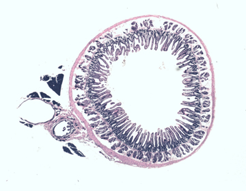
 Color Histology Images
Color (8-bit) gif files of light microscopic images are archived according to tissue type and organ systems. The majority of these are stained with hematoxylin and eosin but in several cases more specialized stains are employed. Index pages and files can be accessed by clicking on red button above.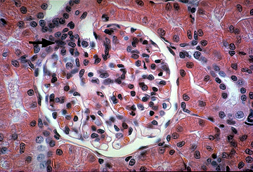
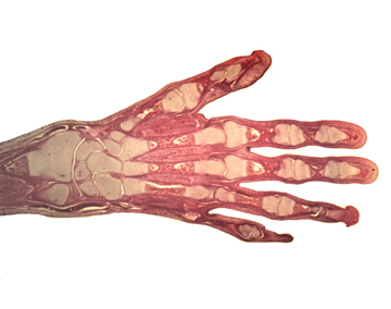
Low Magnification Histology Images
Low magnification images taken with a Leitz Photomakroskope permit surveillance of large tissue regions and entire organs giving a perspective
seldom acheived with a conventional compound microscope.
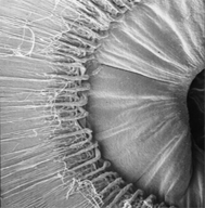
Cell and Tissue Ultrastructure
Transmission and scanning electron micrographs are archived as 8 bit grey-scale images and are listed according to tissue type and organ system. These are meant to compliment the light microscopic images and reveal structural detail not observable by light microscopy.
Mammalian Histology Lectures
Power point presentations and digitized images of labeled illustrations used in lectures are listed according to lecture schedule and can be browsed for study purposes.
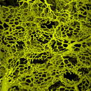
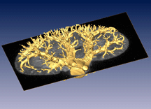
Computer Modeling Of Vascular Corrosion Casts
Blood vascular systems can be injected with a rapidly-hardening polymer. Subsequent corrosion of the surrounding tissue with alkali results in a cast of the blood-space of the original vascular system down to the level of capillaries. These can be examined with light microscopy but the depth of focus is limiting. Scanning electron microscopy of vascular casts results is strikingly sharp images of casts which are in focus throughout their depth. However these can be viewed from only limited angle of tilt and interior casted vessesl are often hidden. The casting plastic is fluorescent and three-dimensional data sets can be acquired with a confocal microscope. These Z-stacks can be thresholded and surface-rendered resulting in a model of the original cast which can be rotated and viewed from any angle. In addition, when the voxel size is calibrated, precise quantitative measurements such as volume, surface area, vessel length and intersect ponts can be calculated. Confocal microscopy can aquired three dimensional data from only very small volumes. Models of whole organ casts can be rendered from Micro-CT scans of casts made from plastic resin plus a lead compound. High resolution Nano-CT scans reveal vascular cast structure to the level of capillaries comparable to Confocal microscopy .
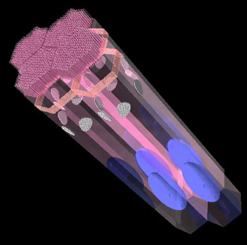
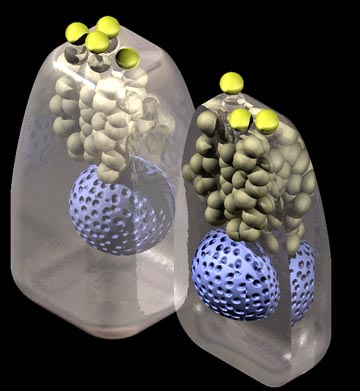
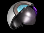
Three Dimensional Models
Three dimensional models of tissue and cell structures have been constructed and rendered to provide realistic interpretations of volume structures not evident in two dimensional tissue sections.
Animated Sequences of Three Dimensional Biological Models
Three-dimensional biological models generated with various versions of STRATAVISION are examined by video sequences of rotating models or camera flythroughs, in and around models.
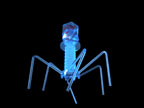
Just for Fun (Wagart Page)- Aliens, Models, Anaglyphs, Colored EMs,Confocals and Other Worlds
-------------------------------------------------------------------------------------------------------------------------------------------------------------
More information for Histology !
http://www.med.umich.edu/histology/cellsTissue/muscle.html
http://www.bu.edu/histology/m/index.htm
http://www.wou.edu/~lemastm/Teaching/BI234/Laboratory%201%20-%20Histology.pdf
http://www.histology-world.com/quizlinks/connective.htm
http://www.udel.edu/biology/Wags/histopage/colorpage/colorpage.htm
http://www.udel.edu/biology/Wags/histopage/modelspage/modelspage.htm
http://www.udel.edu/biology/Wags/histopage/moviespage/moviespage.htm
http://www.udel.edu/biology/Wags/histopage/wagnerart/wagart.html
http://www.courseweb.uottawa.ca/medicine-histology/english/ss_basictissues/connective_tissue.htm
Questions :
http://www.cytochemistry.net/cell-biology/medical/practice_exam_connective_tissue.htm
http://iws.ccccd.edu/rstlaurent/Documents/BIOL_2401_Ex_6A.pdf
http://avalon.unomaha.edu/hpa/2740test2.html


ADIPOSE TISSUE.ppt
