Histology 2
Instructions: In order to use this histology tissue classfication activity, you need a browser that supports flash. Make sure the sound to your computer is on. You will be shown various tissues or organs. Classify each one according to one of the four basic tissue types. Drag it to the appropriate tissue type. Please note: if you are shown an organ, match it up to the basic tissue type which forms the parenchyma of that organ. Click start to begin the timer.
Epithelium
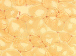
Simple Squamous Epithelium
Histology slide of simple squamous epithelium with audio narration
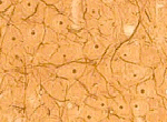
Simple Cuboidal Epithelium
Histology slide of simple cuboidal epithelium with audio narration
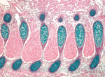
Simple Columnar Epithelium 1 (2)
Histology slide of simple columnar epithelium with audio narration
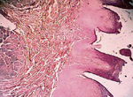
Stratified Squamous Epithelium
Histology slide of stratified squamous epithelium with audio narration
Pseudostratifed Columnar Epithelium
Histology slide of pseudostratifed columnar epithelium with audio narration
Transitional Epithelium
Histology slide of transitional epithelium with audio narration
Connective Tissue
Dense Regular Connective Tissue
Histology slide of dense regular connective tissue with audio narration
Adipose Tissue
Histology slide of adipose tissue with audio narration
Muscle Tissue
Cardiac Muscle
Histology slide of cardiac muscle with audio narration
Skeletal Muscle 1
Histology slide of skeletal muscle with audio narration
Skeletal Muscle 2
Histology slide of skeletal muscle with audio narration
Smooth Muscle
Histology slide of smooth muscle with audio narration
Cartilage
Hyaline Cartilage
Histology slide of hyaline cartilage with audio narration
Fibrocartilage
Histology slide of fibrocartilage with audio narration
Skeletal System
Tendon
Histology slide of tendon with audio narration
Bone
Histology slide of bone with audio narration
Bone (immature)
Histology slide of bone with audio narration
Lymphatic System
Spleen
Histology slide of the spleen with audio narration
Urinary System
Kidney
Histology slide of the kidney with audio narration
Ureter
Histology slide of the ureter with audio narration
Bladder
Histology slide of the bladder with audio narration
Cardiovascular System and Hematopoietic System
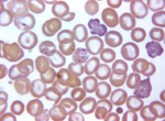
Blood
Histology slide of blood with audio narration
Bone Marrow
Histology slide of bone marrow with audio narration
Blood Vessels
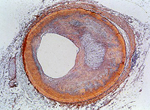
Histology slide of blood vessels with audio narration
Cardiac Muscle
Histology slide of cardiac muscle with audio narration
Respiratory System
Trachea
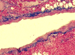
Histology slide of the trachea with audio narration
Bronchus
Histology slide of the bronchus with audio narration
Lung
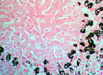
Histology slide of the lungs with audio narration
Gastrointestinal System
Tongue
Histology slide of the tongue with audio narration
Sublingual Gland
Histology slide of the sublingual gland with audio narration
Parotid Gland
Histology slide of the parotid gland with audio narration
Esophagus
Histology slide of the esophagus with audio narration
Stomach
Histology slide of the stomach with audio narration
Small Intestine
Histology slide of the small intestine with audio narration
Large Intestine
Histology slide the large intestine with audio narration
Appendix
Histology slide of the appendix with audio narration
Liver
Histology slide of the liver with audio narration
Gallbladder
Histology slide of the gallbladder with audio narration
Pancreas
Histology slide of the pancreas with audio narration
Integumentary System
Skin
Histology slide of skin with audio narration
Sebacious Gland
Histology slide of sebacious gland with audio narration
Sweat Gland
Histology slide of sweat glands with audio narration
Endocrine System
Adrenal Gland
Histology slide of the adrenal gland with audio narration
Pancreas
Histology slide of the pancreas with audio narration
Pituitary Gland
Histology slide of the pituitary gland with audio narration
Thyroid Gland
Histology slide of the thyroid gland with audio narration
Reproductive System
Ovary
Histology slide of the ovary with audio narration
Seminiferous Tubules
Histology slideof the seminiferous tubules with audio narration
Nervous System and Special Senses
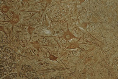
Neurons
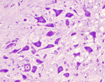
Histology slides of neurons with audio narration
Peripheral Nerve
Histology slide of a peripheral nerves with audio narration
Neuromuscular Junction
Histology slide of the neuromuscular junction with audio narration
Neuroglia and Astrocytes
Histology slide of neuroglia and astrocytes with audio narration
Retina
Histology slide of the retina with audio narration
Cochlea
Histology slide of the cochlea with audio narration
Practical Exam:
|
CellsEpithelium HistologyConnective Tissue HistologyCartilage and Bone HistologyBlood and HematopoiesisMuscle HistologyNervous System HistologyLymphatic System HistologyIntegumentary System HistologyComprehensive Histology 1
Note: If you encoutner a problem with taking the practice practical, make sure you turn on JavaScript. Active Content must likewise be enabled. |
|
Instructions: In order to use the histology flashcards, you need a browser that supports flash. Make sure the sound to your computer is on. Identify the tissue on each flashcard. Click on the flashcard for the answer.
Histology images may be copyrighted and can not be reproduced without permission. Histology images courtesy of anonymous donation, Education Interactive Human Histology Photo CD, Florida State University, and Histology-World.
|
Microscopes test 1, test 2
Histology tests about the microscope
www.histology-world.com/testbank/testbank.htm
Histology Stains and Techniques test 1, test 2
Histology tests about histotechniques
www.histology-world.com/testbank/testbank.htm
Cells test 1, test 2, test 3
Histology tests about cells
www.histology-world.com/testbank/testbank.htm
Histology of Epithelium test 1, test 2, test 3, test 4
Histology tests about epithelium
www.histology-world.com/testbank/testbank.htm
Histology of Connective Tissue test 1, test 2, test 3,test 4
Histology tests about connective tissue
www.histology-world.com/testbank/testbank.htm
Histology of Cartilage test 1, test 2, test 3
Histology tests about cartilage
www.histology-world.com/testbank/testbank.htm
Histology of Bone test 1, test 2, test 3, test 4
Histology tests about bone
www.histology-world.com/testbank/testbank.htm
Histology of Muscle test 1, test 2, test 3, test 4, test 5, test 6
Histology tests about muscle
www.histology-world.com/testbank/testbank.htm
Histology of the Integumentary System test 1, test 2, test 3, test 4, test 5
Histology tests about the integumentary system
www.histology-world.com/testbank/testbank.htm
Histology of the GI System test 1, test 2, test 3,test 4, test 5
Histology tests about the gastrointestinal system
www.histology-world.com/testbank/testbank.htm
Histology of the Pancreas and Hepatobiliary System test 1, test 2, test 3
Histology tests about the hepatobiliary system
www.histology-world.com/testbank/testbank.htm
Histology of the Nervous System test 1, test 2, test 3, test 4, test 5, test 6
Histology tests about the nervous system
www.histology-world.com/testbank/testbank.htm
Histology of the Special Senses test 1, test 2, test 3,test 4, test 5, test 6
Histology tests about the special senses (eye and ear)
www.histology-world.com/testbank/testbank.htm
Volume II
Histology of the Urinary System test 1, test 2, test 3
Histology tests about the urinary system
www.histology-world.com/testbank/testbank.htm
Histology of Blood test 1, test 2, test 3
Histology tests about blood
www.histology-world.com/testbank/testbank.htm
Histology of the Blood Vessels test 1, test 2, test 3,test 4
Histology tests about the blood vessels
www.histology-world.com/testbank/testbank.htm
Histology of the Cardiovascular System test 1
Histology tests about the cardiovascular system
www.histology-world.com/testbank/testbank.htm
Histology of the Lymphatic System test 1, test 2,test 3
Histology tests about the lymphatic system
www.histology-world.com/testbank/testbank.htm
Histology of the Respiratory System test 1, test 2,test 3, test 4
Histology tests about the respiratory system
www.histology-world.com/testbank/testbank.htm
Histology of the Endocrine System test 1, 2, 3, 4, 5,6, 7
Histology tests about the endocrine system
www.histology-world.com/testbank/testbank.htm
Histology of the Male Reproductive System test 1,test 2, test 3
Histology tests about the male reproductive system
www.histology-world.com/testbank/testbank.htm
Histology of the Female Reproductive System test 1,test 2
Histology tests about the female reproductive system
www.histology-world.com/testbank/testbank.htm
nsructions: When the histology quiz question is asked, click on the button for the correct answer. Have your histology book handy! When you are ready for the next histology quiz question, click the "next question" button.
Histology Quiz Master 1 Histology Quiz Master 2
|
Aldehyde Fuchsin Alician Blue
Alizarin Red S Alkaline Phosphatase
Bielschowsky Stain
Cajal Stain
Congo Red
Cresyl Violet
Fontana-Masson
Giemsa Stain This micrograph depicts the appearance of a well-stained slide using the Giemsa staining technique.
Golgi Stain
Gomori Trichrome H&E Hematoxylin can be thought of as a basic dye. It binds to acidic structures, staining them blue to purple. It will bind and stain nucleic acids. Therefore, the nucleus stains blue. Eosin is an acid aniline dye. It will bind to and stain basic structures (or negatively charged structures), such as cationic amino groups on proteins. It stains them pink. Cytoplasm, muscle, connective tissue, colloid, red blood cells and decalcified bone matrix all stain pink to pink/orange/red with eosin. With an H&E stain, mucus and cartilage will stain a light blue color.
Iron Hematoxylin Luna Stain
Luxol Fast Blue
Mallory Trichome Masson Trichome Collagen fibers stain green or blue with Masson's trichrome stain. Muscle and keratin will be red. Cytoplasm will be pink to red. Nuclei will be black.
Movat's Pentachrome Stain
Mucicarmine Nissl Stains
Nuclear Fast Red
Oil Red O
Orcien Stain Osmium Tetroxide
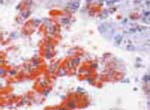
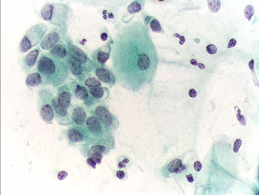
Papanicolaou Stain
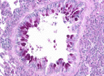
Periodic Acid-Schiff (PAS)
Phosphotungstic Acid-Hematoxylin (PTAH)
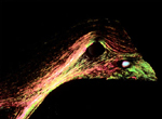
PicroSirius Red (polarized)
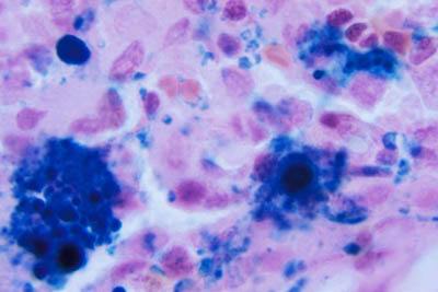
Prussian Blue
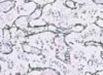
Reticular Fiber Stain
Romanowsky Stains Safranin O
Silver Stains Sudan Stains Tartrazine Toludine Blue
Van Gieson Verhoeff Stain
Von Kossa Stain
Wright's Stain This histology stain uses a blend of basic dyes, such as methylene blue derivatives and acid dyes, such as eosin. Red blood cells stain reddish/pink. Eosinophilic granules will be a bright orange/red. The nucleus of white blood cells will stain purple. Basophilic granules will stain blue/black. Neutrophilic granules stain pale brown. Platelets stain purple. The cytoplasm of lymphocytes stains pale blue. This histology slide has been stained using an "automatic staining" device. It is a Wright's stain. The acidic components, such as the nucleic acids, will take on the basic portion of the stain, or the methylene azure color, and stain blue. The basic components, such as erythrocyte hemoglobin, will stain pink with eosin, since eoisn is an acidic dye.
|
|
GPA {Growth hormone and Prolactin are secreted by the Acidophilic cells of the anterior pituitary} |
|
Never Let Momma Eat Beans {neutrophils, lymphocytes, monocytes, eosinophils, basophils} |
|
|
|
Never Let Monkeys Eat Bananas {neutrophils, lymphocytes, monocytes, eosinophils, basophils}
|
Canadian Ladies Give Superb Backrubs
{corneum, lucidum, granulosum, spinosum, basale}
 |
The deeper you go, the sweeter it gets: salt, sugar, sex! {the order of the products of the adrenal cortex: mineralocorticoids, glucocorticoids, androgens} |
Go Find Rex
Make Good Sex!
{zona glomerulosa, zona fasciculata, zona retiuclaris
mineralocorticoid, glucocorticoid, sex steroid}
 |
Worms, wheezes, weird diseases {eosinophilia} |
B-FLAT
{the Basophilic cells of the anterior pituitary secreteFSH, LH, ACTH, TSH}
